
Prions
in the Ocean: A Natural Case of Prion Disease in Dolphins
Manuel Camacho,1,† Diego Morales-Schehing,1, 2 Natalia Fernandez-Borges,3
Joaquin Castilla,1, 3 Daniel Cowan4 and Claudio Soto,1
1Department of Neurology, University of Texas Houston Medical School;
Houston, TX USA; 2Facultad de Medicina, Universidad de Los Andes; Santiago,
Chile; 3CIC bioGUNE & IKERBASQUE, Basque Foundation for Science; Bilbao,
Spain; 4Department of Pathology, University of Texas Medical Branch; Houston, TX
USA †Presenting author; Email: Manuel.V.CamahoMartinez@uth.tmc.edu
Prion diseases or Transmissible Spongiform Encephalopathies (TSEs) are
neurodegenerative disorders associated with the misfolding of the prion protein
(PrPSc). TSEs are known to affect naturally various species of mammals,
including humans, cattle, sheep, goats, cervids, felines and mink. In this study
we report the first potential case of TSEs in marine mammals. Dolphin (Tursiops
truncatus) has a relatively long lifespan and a complex brain structure.
Moreover, the PrP sequence has a large similarity with those of other mammal
species naturally affected by TSEs. Fortuitously we identified a dolphin
exhibiting behavioral alterations evidently suggestive of a brain disease. These
alterations included aggressiveness, lack of motor coordination and anti-social
behavior. An initial examination of the brain stained by hematoxilin/eosin
showed extensive spongiform degeneration, especially in areas of the cortex and
cerebellum. We subsequently analyzed brain tissue from this animal and compared
with staining from various healthy dolphins by immunohistochemistry using the
6H4 antibody that recognizes the amino acid residues 144-152 of the PrP
sequence, highly conserved in most mammalian species. The result showed spread
diffuse staining with some more punctuated immune-reactivity, reminiscent of
amyloid plaques. This staining was substantially different from the light
background reactivity of healthy brains. In addition, an anti-GFAP antibody was
used in brain slices in order to assess a possible inflammatory response, which
is commonly associated to prion diseases. The results clearly showed profuse
astrogliosis, especially in the areas close to PrP deposition. Biochemical
studies showed the presence of PK resistant forms of PrP with a shift in their
electrophoretical mobility, similar to what is found in natural and
experimentally induced TSEs. Additionally, in vitro experiments showed that
dolphin PrPC can be converted into the PrPSc form by PMCA using prions from
various species. We are currently sequencing the prnp gene in this animal to
identify any possible mutation. Our findings show for the first time a
prion-like disease in a marine mammal, indicating that this intriguing
neurodegenerative disorder could be present in a wide spectrum of species, even
under the sea.
Prion biology - Landes Bioscience Home
JOURNAL OF BIOLOGICAL REGULATORS & HOMEOSTATIC AGENTS
LETTER TO THE EDITOR
PRION SEARCH AND CELLULAR PRION PROTEIN EXPRESSION IN STRANDED DOLPHINS
SC/65a/Forinfo04 Vo\. 26, no. 3, 567-570 (2012)
G. DI GUARDOl, C. COCUMELLP, R. MEOLP, K. BARBAR02, G. TERRACCIAN03, C.E.
DI FRANCESCO!, S. MAZZARIOL4 and C. ELENP 'Department oj Comparative Biomedical
Sciences, University ojTeramo, Teramo, Italy; 2Istituto Zooprojilattico
Sperimentale (IZS) delle Regioni Lazio e Toscana, Rome, Italy; 3IZS delle
Regioni Lazio e Toscana, Pisa, Italy; "Department of Comparative Biomedicine and
Food Science, University ojPadova, AGRIPOLIS, Legnaro, Padua, Italy Received May
18, 2012 - Accepted July 18, 2012
The recent description of a prion disease (PD) case in a free-ranging
bottlenose dolphin (Tursiops truncatus) prompted us to carry out an extensive
search for the "disease-associated" isoform (PrPSC) of the cellular prion
protein (PrPC) in the brain and in a range of lymphoid tissues from 23 striped
dolphins (Stenella coeruleoalba), 5 bottlenose dolphins and 2 Risso's dolphins
(Grampus griseus) found stranded between 2007 and 2012 along the Italian
coastline. Three striped dolphins and one bottlenose dolphin showed microscopic
lesions of encephalitis, with no evidence of spongiform brain lesions being
detected in any ofthe 30 free-ranging cetaceans investigated herein.
Nevertheless, we could still observe a prominent Pr'P" immunoreactivity in the
brain as well as in lymphoid tissues from these dolphins. Although
immunohistochemical and Western blot investigations yielded negative results for
Prpsc deposition in all tissues from the dolphins under study, the reported
occurrence of a spontaneous PD case in a wild dolphin is an intriguing issue and
a matter of concern for both prion biology and intra/ inter-species
transmissibility, as well as for cetacean conservation medicine.
snip...
Secondly, on the basis of the high sequence homology between cetacean PRNP
gene and that of the aforementioned ungulate species, which may naturally
develop bovine spongiform encephalopathy (BSE) and scrapie (4), susceptibility
to and occurrence of TSEs in cetaceans have been deemed plausible (2).
Additionally, one of the most, if not even the most intriguing feature of a TSE-
like condition in a free-living cetacean refers to the route(s) through which
such prion infection may have been acquired by the animal under study (1).
snip...
As previously mentioned, one of the most, if not even the most challenging
feature of a PD condition in free-living cetaceans refers to the route(s)
through which infection may be acquired. In this respect, whenever a "sporadic"
form of PD developed in a dolphin, similarly to what has been known for over 90
years in humans (4), this would probably lead to a different "scenario" from
that which we would face in the case of wild dolphins being found to be
susceptible to an "infectious" PD condition, as in the well-documented example
of sheep scrapie (4). In fact, this would represent an additional threat to
cetaceans, the health and conservation status of which are dramatically impacted
nowadays by many other biological and anthropogenic noxae (10). With reference
to the above, further issues of concern regard the animal source(s) of infection
and its putative "land-to-sea" flow, as already hypothesized for Toxoplasma
gondii infection in sea otters (En hydra lutris) and bottlenose dolphins (10),
or the alternative existence of an "exclusively marine" cycle of infection.
Finally, a particularly relevant issue is that addressing the "strategies"
adopted by dolphin prions for their persistence within the marine environment,
considering their progressive dilution and dispersal in sea water.
In conclusion, despite the negative results of our study, we still believe
that the documented occurrence of a spontaneous TSE-like condition in a wild
dolphin (1) is an intriguing issue and a matter of concern for both prion
biology and intra/inter-species transmissibility, as well as for cetacean
conservation medicine.
Mol Psychiatry. 1997 Mar;2(2):146-7. Normal isoform of amyloid protein
(PrP) in brains of spawning salmon. Gibbs CJ Jr, Bolis CL. Source Laboratory of
Central Nervous System Studies, National Institute of Neurological Disorders and
Stroke, National Institutes of Health, Bethesda, MD, USA.
Abstract The transmissible spongiform encephalopathies are a group of
subacute progressive degenerative diseases of the nervous system which are
always fatal in their outcome. These diseases appear to be caused by the
abnormal isoform of the precursor protein of amyloid designated prion protein.
The normal isoform has been identified in the tissues of all mammalian species
thus far tested as well as in Drosophila. We report the presence of this protein
for the first time in the brains of fish.
Greeetings mad cow tse prion world,
disturbing and interesting to say the least, but not surprising, dolphin
are mammals. now, what type TSE prions is man putting in the water to uptake,
dare I ask ???
FIRST, what about our beaches, and how are hospitals disposing of their
waste ???

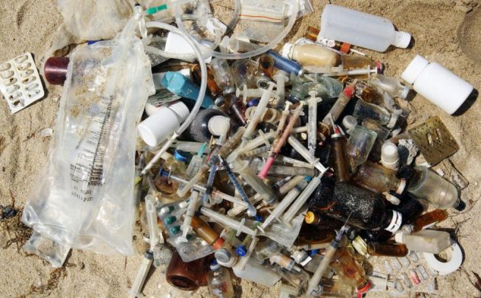
how about mortuaries and their blood and waste ???
Baris Funeral Home, Clyde NY, Wayne County – Embalming
STEP 3- Embalming Process
Incisions are made in both vessels, and a tube connected to the the
embalming fluid pump is placed into the carotid artery, Another tube is placed
into the jugular vein, this is called a drain tube. The basic theory is to pump
embalming fluid into the artery, and this will cause the blood to return through
the veins and flow outside the remains for disposal. Approximately 3 gallons of
a mixture of fluid and water are circulated through the remains for thorough
disinfection and preservation to take place. In most cases, this will be the
only point of injection ofthe embalming fluid. There are times when clots and
other factors stop the flow of fluid throughout out the whole system, and at
these times, other points of injection are necessary in order to do a complete
and thorough embalming. There are many factors which go into the process, which
cannot be explained here due to space limitations, but some of the factors that
the funeral director must assess before embalming are the mode of death, the
weight of the remains, the general overall condition of the remains, any disease
associated with the remains, etc. These factors determine the types and
strengths of fluids used, and the type of embalming necessary to complete the
task. Many fluids have a slight dye added to them, which gives the remains a
pinkish glow, and also acts as a guide for the funeral director, making it
visible for him to see the fluid as it travels through the remains. This type of
embalming is known as arterial embalming.
A commonly asked question at this point is:
What do you do with the blood you remove from the body?
Once the blood mixes with the embalming chemicals, it becomes basically
harmless.
The laws allow us to put the blood down the normal sanitary sewer drains in
the preparation room sink as it does not pose a health risk.
what about sludge ???
Envt.04: Detection of Prions in Anaerobic Digestion Sludge by PMCA
Shannon Braithwaite,3,† Brandon H. Gilroyed,2 Tim Reuter,4 Stefanie Czub,5
Catherine Graham,5 Aru Balachandran,6 Tim McAllister,2 Miodrag Belosevic7, 3 and
Norman F. Neumann3, 8
1University of Alberta; Edmonton, AB Canada; 2Agriculture and Agri-Food
Canada; Lethbridge, AB Canada; 3 School of Public Health, University of Alberta;
Edmonton, AB Canada; 4Alberta Agriculture and Rural Development; Lethbridge, AB
Canada; 5Canadian Food Inspection Agency; Lethbridge, AB Canada; 6Canadian Food
Inspection Agency; Ottawa, ON Canada; 7Department of Biological Sciences,
University of Alberta; Edmonton, AB Canada; 8Alberta Provincial Laboratory for
Public Health; Edmonton, AB Canada;
†Presenting author; Email: shannon.braithwaite@ualberta.ca
The exceptional physio-chemical resistance of prions to established
decontamination procedures poses a challenge to assess the suitability of
applied inactivation approaches. The capacity of prion detection is limited by
the sensitivity level of western blotting (WB) or by the cost and time factors
of bioassays. In addition, prion detection assays might be limited by either the
unique or complex nature of matrices associated with environmental samples. We
investigated anaerobic digestion (AD) as a practical and economic approach for
potential re-cycling of specified risk material (SRM) into value added products
(i.e., renewable energy). However, the complex matrix of AD poses a challenge to
verify prion detection and degradation. The sensitivity of protein misfolding
cyclic amplification (PMCA) with subsequent WB visualization, offers a
sensitivity level required for the evaluation of prion biodegradation
strategies. AD sludge inhibited the PMCA reaction and/or western blot detection.
However, at concentrations less than <1 263k="" a="" ad="" amplified="" anaerobic="" and="" area="" be="" bench-scale="" bioavailable="" biological="" complex="" concentrations="" could="" currently="" degradation="" detected.="" digester="" div="" experiment="" fate="" for="" high="" in="" inactivation.="" infectious="" insight="" into="" investigate="" is="" l="" matrices.="" of="" or="" potential="" presence="" present="" prion="" prions="" proven="" provide="" research="" scrapie="" semi-quantitatively="" sludge="" the="" to="" underway="" valuable="" were="" will="">
A Quantitative Assessment of the Amount of Prion Diverted to Category 1
Materials and Wastewater During Processing
Amie Adkin1,*, Neil Donaldson1, Louise Kelly1,2Article first published
online: 24 DEC 2012
DOI: 10.1111/j.1539-6924.2012.01922.x
© 2012 Society for Risk Analysis
Keywords:Abattoir;bovine spongiform encephalopathy;QRA;scrapie;TSE
In this article the development and parameterization of a quantitative
assessment is described that estimates the amount of TSE infectivity that is
present in a whole animal carcass (bovine spongiform encephalopathy [BSE] for
cattle and classical/atypical scrapie for sheep and lambs) and the amounts that
subsequently fall to the floor during processing at facilities that handle
specified risk material (SRM). BSE in cattle was found to contain the most oral
doses, with a mean of 9864 BO ID50s (310, 38840) in a whole carcass compared to
a mean of 1851 OO ID50s (600, 4070) and 614 OO ID50s (155, 1509) for a sheep
infected with classical and atypical scrapie, respectively. Lambs contained the
least infectivity with a mean of 251 OO ID50s (83, 548) for classical scrapie
and 1 OO ID50s (0.2, 2) for atypical scrapie. The highest amounts of infectivity
falling to the floor and entering the drains from slaughtering a whole carcass
at SRM facilities were found to be from cattle infected with BSE at rendering
and large incineration facilities with 7.4 BO ID50s (0.1, 29), intermediate
plants and small incinerators with a mean of 4.5 BO ID50s (0.1, 18), and
collection centers, 3.6 BO ID50s (0.1, 14). The lowest amounts entering drains
are from lambs infected with classical and atypical scrapie at intermediate
plants and atypical scrapie at collection centers with a mean of 3 × 10⁻⁷ OO
ID50s (2 × 10⁻⁸, 1 × 10⁻⁶) per carcass. The results of this model provide key
inputs for the model in the companion paper published here.
We also detected PrP(CWD) in one of two environmental water samples from a
CWD endemic area collected at a time of increased water runoff from melting
winter snow pack, as well as in water samples obtained concurrently from the
flocculation stage of water processing by the municipal water treatment
facility.
Detection of Protease-Resistant Prion Protein in Water from a CWD-Endemic
Area
65
Tracy A. Nichols*1,2, Bruce Pulford1, Christy Wyckoff1,2, Crystal
Meyerett1, Brady Michel1, Kevin Gertig3, Jean E. Jewell4, Glenn C. Telling5 and
M.D. Zabel1 1Department of Microbiology, Immunology and Pathology, College of
Veterinary Medicine and Biomedical Sciences, Colorado State University, Fort
Collins, CO 80523, USA 2National Wildlife Research Center, Wildlife Services,
United States Department of Agriculture, Fort Collins, Colorado, 80521, USA
3Fort Collins Water and Treatment Operations, Fort Collins, Colorado, 80521, USA
4 Department of Veterinary Sciences, Wyoming State Veterinary Laboratory,
University of Wyoming, Laramie, Wyoming, 82070, USA 5Department of Microbiology,
Immunology, Molecular Genetics and Neurology, Sanders Brown Center on Aging,
University of Kentucky, Lexington, Kentucky, 40536, USA * Corresponding author-
tracy.a.nichols@aphis.usda.gov
Chronic wasting disease (CWD) is the only known transmissible spongiform
encephalopathy affecting free-ranging wildlife. Experimental and epidemiological
data indicate that CWD can be transmitted horizontally and via blood and saliva,
although the exact mode of natural transmission remains unknown. Substantial
evidence suggests that prions can persist in the environment, implicating it as
a potential prion reservoir and transmission vehicle. CWD- positive animals can
contribute to environmental prion load via biological materials including
saliva, blood, urine and feces, shedding several times their body weight in
possibly infectious excreta in their lifetime, as well as through decomposing
carcasses. Sensitivity limitations of conventional assays hamper evaluation of
environmental prion loads in water. Here we show the ability of serial protein
misfolding cyclic amplification (sPMCA) to amplify minute amounts of CWD prions
in spiked water samples at a 1:1 x10⁶ , and protease-resistant prions in
environmental and municipal-processing water samples from a CWD endemic area.
Detection of CWD prions correlated with increased total organic carbon in water
runoff from melting winter snowpack. These data suggest prolonged persistence
and accumulation of prions in the environment that may promote CWD transmission.
snip...
The data presented here demonstrate that sPMCA can detect low levels of
PrPCWD in the environment, corroborate previous biological and experimental data
suggesting long term persistence of prions in the environment2,3 and imply that
PrPCWD accumulation over time may contribute to transmission of CWD in areas
where it has been endemic for decades. This work demonstrates the utility of
sPMCA to evaluate other environmental water sources for PrPCWD, including
smaller bodies of water such as vernal pools and wallows, where large numbers of
cervids congregate and into which prions from infected animals may be shed and
concentrated to infectious levels.
snip...end...full text at ;
We also detected PrP(CWD) in one of two environmental water samples from a
CWD endemic area collected at a time of increased water runoff from melting
winter snow pack, as well as in water samples obtained concurrently from the
flocculation stage of water processing by the municipal water treatment
facility.
AS the crow flies, so to the TSE prion disease
Sunday, July 07, 2013
Could avian scavengers translocate infectious prions to disease-free areas
initiating new foci of chronic wasting disease?
Prion. 2013 Jul 3;7(4). [Epub ahead of print]
Wednesday, July 10, 2013
Rapid assessment of bovine spongiform encephalopathy prion inactivation by
heat treatment in yellow grease produced in the industrial manufacturing process
of meat and bone meals
BMC Veterinary Research 2013, 9:134 doi:10.1186/1746-6148-9-134
PLoS ONE. 2008; 3(8): e2969.
Prion Infected Meat-and-Bone Meal Is Still Infectious after Biodiesel
Production
The epidemic of bovine spongiform encephalopathy (BSE) has led to a
world-wide drop in the market for beef by-products, such as Meat-and-Bone Meal
(MBM), a fat-containing but mainly proteinaceaous product traditionally used as
an animal feed supplement. While normal rendering is insufficient, the
production of biodiesel from MBM has been suggested to destroy infectivity from
transmissible spongiform encephalopathies (TSEs). In addition to producing fuel,
this method simultaneously generates a nutritious solid residue. In our study we
produced biodiesel from MBM under defined conditions using a modified form of
alkaline methanolysis. We evaluated the presence of prion in the three resulting
phases of the biodiesel reaction (Biodiesel, Glycerol and Solid Residue) in
vitro and in vivo. Analysis of the reaction products from 263K scrapie infected
MBM led to no detectable immunoreactivity by Western Blot. Importantly, and in
contrast to the biochemical results the solid MBM residue from the reaction
retained infectivity when tested in an animal bioassay. Histochemical analysis
of hamster brains inoculated with the solid residue showed typical spongiform
degeneration and vacuolation. Re-inoculation of these brains into a new cohort
of hamsters led to onset of clinical scrapie symptoms within 75 days, suggesting
that the specific infectivity of the prion protein was not changed during the
biodiesel process. The biodiesel reaction cannot be considered a viable prion
decontamination method for MBM, although we observed increased survival time of
hamsters and reduced infectivity greater than 6 log orders in the solid MBM
residue. Furthermore, results from our study compare for the first time prion
detection by Western Blot versus an infectivity bioassay for analysis of
biodiesel reaction products. We could show that biochemical analysis alone is
insufficient for detection of prion infectivity after a biodiesel process.
New studies on the heat resistance of hamster-adapted scrapie agent:
Threshold survival after ashing at 600°C suggests an inorganic template of
replication
Abstract
One-gram samples from a pool of crude brain tissue from hamsters infected
with the 263K strain of hamster-adapted scrapie agent were placed in covered
quartz-glass crucibles and exposed for either 5 or 15 min to dry heat at
temperatures ranging from 150°C to 1,000°C. Residual infectivity in the treated
samples was assayed by the intracerebral inoculation of dilution series into
healthy weanling hamsters, which were observed for 10 months; disease
transmissions were verified by Western blot testing for proteinase-resistant
protein in brains from clinically positive hamsters. Unheated control tissue
contained 9.9 log10LD50/g tissue; after exposure to 150°C, titers equaled or
exceeded 6 log10LD50/g, and after exposure to 300°C, titers equaled or exceeded
4 log10LD50/g. Exposure to 600°C completely ashed the brain samples, which, when
reconstituted with saline to their original weights, transmitted disease to 5 of
35 inoculated hamsters. No transmissions occurred after exposure to 1,000°C.
see full text:
PRODUCT Bulk custom dairy pre-mixes,
Recall # V-120-6 CODE None RECALLING FIRM/MANUFACTURER Ware Milling Inc.,
Houston, MS, by telephone on June 23, 2006. Firm initiated recall is complete.
REASON Possible contamination of dairy animal feeds with ruminant derived meat
and bone meal.
VOLUME OF PRODUCT IN COMMERCE 350 tons
DISTRIBUTION AL and MS
______________________________
PRODUCT
*** a) Tucker Milling, LLC Tm 32% Sinking Fish Grower, #2680-Pellet, 50 lb.
bags, Recall # V-121-6;
b) Tucker Milling, LLC #31120, Game Bird Breeder Pellet, 50 lb. bags,
Recall # V-122-6;
c) Tucker Milling, LLC #31232 Game Bird Grower, 50 lb. bags, Recall #
V-123-6;
d) Tucker Milling, LLC 31227-Crumble, Game Bird Starter, BMD Medicated, 50
lb bags, Recall # V-124-6;
e) Tucker Milling, LLC #31120, Game Bird Breeder, 50 lb bags, Recall #
V-125-6;
f) Tucker Milling, LLC #30230, 30 % Turkey Starter, 50 lb bags, Recall #
V-126-6;
g) Tucker Milling, LLC #30116, TM Broiler Finisher, 50 lb bags, Recall #
V-127-6
CODE All products manufactured from 02/01/2005 until 06/20/2006 RECALLING
FIRM/MANUFACTURER Recalling Firm: Tucker Milling LLC, Guntersville, AL, by
telephone and visit on June 20, 2006, and by letter on June 23, 2006.
Manufacturer: H. J. Baker and Brothers Inc., Stamford, CT. Firm initiated recall
is ongoing.
REASON Poultry and fish feeds which were possibly contaminated with
ruminant based protein were not labeled as "Do not feed to ruminants".
VOLUME OF PRODUCT IN COMMERCE 7,541-50 lb bags
DISTRIBUTION AL, GA, MS, and TN
END OF ENFORCEMENT REPORT FOR AUGUST 9, 2006
###
Subject: MAD COW FEED RECALL AL AND FL VOLUME OF PRODUCT IN COMMERCE 125
TONS Products manufactured from 02/01/2005 until 06/06/2006
Date: August 6, 2006 at 6:16 pm PST PRODUCT
*** a) CO-OP 32% Sinking Catfish, Recall # V-100-6;
b) Performance Sheep Pell W/Decox/A/N, medicated, net wt. 50 lbs, Recall #
V-101-6;
c) Pro 40% Swine Conc Meal -- 50 lb, Recall # V-102-6;
d) CO-OP 32% Sinking Catfish Food Medicated, Recall # V-103-6;
e) "Big Jim's" BBB Deer Ration, Big Buck Blend, Recall # V-104-6;
f) CO-OP 40% Hog Supplement Medicated Pelleted, Tylosin 100 grams/ton, 50
lb. bag, Recall # V-105-6;
g) Pig Starter Pell II, 18% W/MCDX Medicated 282020, Carbadox -- 0.0055%,
Recall # V-106-6;
h) CO-OP STARTER-GROWER CRUMBLES, Complete Feed for Chickens from Hatch to
20 Weeks, Medicated, Bacitracin Methylene Disalicylate, 25 and 50 Lbs, Recall #
V-107-6;
i) CO-OP LAYING PELLETS, Complete Feed for Laying Chickens, Recall # 108-6;
j) CO-OP LAYING CRUMBLES, Recall # V-109-6;
k) CO-OP QUAIL FLIGHT CONDITIONER MEDICATED, net wt 50 Lbs, Recall #
V-110-6;
l) CO-OP QUAIL STARTER MEDICATED, Net Wt. 50 Lbs, Recall # V-111-6;
m) CO-OP QUAIL GROWER MEDICATED, 50 Lbs, Recall # V-112-6 CODE
Product manufactured from 02/01/2005 until 06/06/2006
RECALLING FIRM/MANUFACTURER Alabama Farmers Cooperative, Inc., Decatur, AL,
by telephone, fax, email and visit on June 9, 2006. FDA initiated recall is
complete.
REASON Animal and fish feeds which were possibly contaminated with ruminant
based protein not labeled as "Do not feed to ruminants".
VOLUME OF PRODUCT IN COMMERCE 125 tons
DISTRIBUTION AL and FL
END OF ENFORCEMENT REPORT FOR AUGUST 2, 2006
###
MAD COW FEED RECALL USA EQUALS 10,878.06 TONS NATIONWIDE Sun Jul 16, 2006
09:22 71.248.128.67
RECALLS AND FIELD CORRECTIONS: VETERINARY MEDICINE -- CLASS II
______________________________
PRODUCT
a) PRO-LAK, bulk weight, Protein Concentrate for Lactating Dairy Animals,
Recall # V-079-6;
b) ProAmino II, FOR PREFRESH AND LACTATING COWS, net weight 50lb (22.6 kg),
Recall # V-080-6;
c) PRO-PAK, MARINE & ANIMAL PROTEIN CONCENTRATE FOR USE IN ANIMAL FEED,
Recall # V-081-6;
d) Feather Meal, Recall # V-082-6 CODE
a) Bulk
b) None
c) Bulk
d) Bulk
RECALLING FIRM/MANUFACTURER H. J. Baker & Bro., Inc., Albertville, AL,
by telephone on June 15, 2006 and by press release on June 16, 2006. Firm
initiated recall is ongoing.
REASON
Possible contamination of animal feeds with ruminent derived meat and bone
meal.
VOLUME OF PRODUCT IN COMMERCE 10,878.06 tons
DISTRIBUTION Nationwide
END OF ENFORCEMENT REPORT FOR July 12, 2006
###
Friday, October 8, 2010
Scientific reasons for a feed ban of meat-and-bone meal, applicable to all
farmed animals including cattle, pigs, poultry, farmed fish and pet food
Saturday, November 6, 2010
TAFS1 Position Paper on Position Paper on Relaxation of the Feed Ban in the
EU
Berne, 2010 TAFS INTERNATIONAL FORUM FOR TRANSMISSIBLE ANIMAL DISEASES AND
FOOD SAFETY a non-profit Swiss Foundation
Archive Number 20101206.4364 Published Date 06-DEC-2010 Subject
PRO/AH/EDR>
Prion disease update 2010 (11) PRION DISEASE UPDATE 2010 (11)
P.9.21
Molecular characterization of BSE in Canada
Jianmin Yang1, Sandor Dudas2, Catherine Graham2, Markus Czub3, Tim
McAllister1, Stefanie Czub1 1Agriculture and Agri-Food Canada Research Centre,
Canada; 2National and OIE BSE Reference Laboratory, Canada; 3University of
Calgary, Canada
Background: Three BSE types (classical and two atypical) have been
identified on the basis of molecular characteristics of the misfolded protein
associated with the disease. To date, each of these three types have been
detected in Canadian cattle.
Objectives: This study was conducted to further characterize the 16
Canadian BSE cases based on the biochemical properties of there associated
PrPres. Methods: Immuno-reactivity, molecular weight, glycoform profiles and
relative proteinase K sensitivity of the PrPres from each of the 16 confirmed
Canadian BSE cases was determined using modified Western blot analysis.
Results: Fourteen of the 16 Canadian BSE cases were C type, 1 was H type
and 1 was L type. The Canadian H and L-type BSE cases exhibited size shifts and
changes in glycosylation similar to other atypical BSE cases. PK digestion under
mild and stringent conditions revealed a reduced protease resistance of the
atypical cases compared to the C-type cases. N terminal- specific antibodies
bound to PrPres from H type but not from C or L type. The C-terminal-specific
antibodies resulted in a shift in the glycoform profile and detected a fourth
band in the Canadian H-type BSE.
Discussion: The C, L and H type BSE cases in Canada exhibit molecular
characteristics similar to those described for classical and atypical BSE cases
from Europe and Japan. This supports the theory that the importation of BSE
contaminated feedstuff is the source of C-type BSE in Canada. *It also suggests
a similar cause or source for atypical BSE in these countries.
Saturday, August 4, 2012
*** Final Feed Investigation Summary - California BSE Case - July 2012
What irks many scientists is the USDA’s April 25 statement that the rare
disease is “not generally associated with an animal consuming infected feed.”
The USDA’s conclusion is a “gross oversimplification,” said Dr. Paul Brown,
one of the world’s experts on this type of disease who retired recently from the
National Institutes of Health.
"(The agency) has no foundation on which to base that statement.”
“We can’t say it’s not feed related,” agreed Dr. Linda Detwiler, an
official with the USDA during the Clinton Administration now at Mississippi
State.
In the May 1 email to me, USDA’s Cole backed off a bit. “No one knows the
origins of atypical cases of BSE,” she said
Saturday, May 26, 2012
Are USDA assurances on mad cow case 'gross oversimplification'?
Saturday, December 15, 2012
Bovine spongiform encephalopathy: the effect of oral exposure dose on
attack rate and incubation period in cattle -- an update 5 December 2012
Thursday, June 6, 2013
BSE TSE PRION USDA FDA MAD COW FEED COMPLIANCE REPORT and NAI, OAI, and VAI
ratings as at June 5, 2013
Tuesday, June 11, 2013
Weld County Bi-Products dba Fort Morgan Pet Foods 6/1/12 significant
deviations from requirements in FDA regulations that are intended to reduce the
risk of bovine spongiform encephalopathy (BSE) within the United States
Friday, December 14, 2012
Susceptibility of domestic cats to chronic wasting disease
Saturday, July 6, 2013
Small Ruminant Nor98 Prions Share Biochemical Features with Human
Gerstmann-Sträussler-Scheinker Disease and Variably Protease-Sensitive
Prionopathy
Research Article
Friday, December 14, 2012
Susceptibility Chronic Wasting Disease (CWD) in wild cervids to Humans 2005
- December 14, 2012
Friday, December 14, 2012
DEFRA U.K. What is the risk of Chronic Wasting Disease CWD being introduced
into Great Britain? A Qualitative Risk Assessment October 2012
TSS
No comments:
Post a Comment
Note: Only a member of this blog may post a comment.